Borrelia burgdorferi-specific intrathecal antibody production in neuroborreliosis: A follow-up study.
Susanne Hammers-Berggren, MD; Klaus Hansen, MD; Anne-Mette Lebech, MD; and Mats Karlsson, MD, PhD
Neurology. 1993;43:169-175. PMID: 8423881

Received April , 1992. Accepted for publication in final form June 9, 1992.

OCR, review and comments by Marie Kroun, MD - LymeRICK (english) & DanInfekt & project sitemap (danish)

Article abstract - We used a capture ELISA with biotinylated Borrelia burgdorferi flagella as antigen to analyze the kinetics of intrathecal antibody production against B burgdorferi in 27 patients with neuroborreliosis. All patients had lymphocytic pleocytosis, 13/27 had intrathecal specific IgM production, and 26/27 had intrathecal IgG synthesis against B burgdorferi before therapy. All patients improved after antibiotic treatment. At follow-up, 11 months to 8 years later (median, 114 years), 20 patients had had a complete clinical recovery, and seven suffered from sequelae. One patient without sequelae had persistent specific intrathecal IgM synthesis. Ten of 20 patients without sequelae and five of seven patients with sequelae had persistent intrathecal IgG production against B burgdorferi. None of the 16 patients with persistent specific intrathecal antibody synthesis had pleocytosis at follow-up. Therefore, intrathecal immunoglobulin production against B burgdorferi, especially IgG, may persist for years after treatment of neuroborreliosis without clinical signs of active disease.

Borrelia burgdorferi, the etiologic agent of Lyme borreliosis, is transmitted to humans by Ixodes ticks.1,2
1.Burgdorfer W, Barbour AG, Hayes SF, Benach JL, Grunwaldt E, Davis JP. Lyme disease: a tick borne spirochetosis? Science 1982;216:1317-1319. PMID:  7043737
2. Steere AC, Grodzicki RL, Kornblatt AN, et al. The spirochetal etiology of Lyme disease. N Engi J Med 1983;308:733-740. PMID:   6828118
The spirochetes may spread locally in the skin or disseminate to other organs, most commonly the nervous system, the joints, and the heart.3-7
3. Steere AC, Malawista SE, Snydman DR, et al. Lyme arthritis: an epidemic of oligoarticular arthritis in children and aduits in three Connecticut cominunities. Arthritis Rheum 1977;20:7-17. PMID:
4. Steere AC, Batsford WP, Weinberg M, et al. Lyme carditis: cardiac abnormalities in Lyme disease. Ann Intern Med 1980;93 :8-16. PMID: 836338

5. Steere AC, Bartenhagen NH, Craft JE, et al. The early clinical manifestations of Lyme disease. Ann Intern Med 1983;99:76-82. PMID:  6859726
6. Stiernstedt G. Tick borne Borrelia infection in Sweden. Scand J Infect Dis Suppl 1985;45:40-49. PMID:  3903977
7. Stiernstedt G, Gustavsson R, Karlsson M, Svenungsson B, Sköldenberg B. Clinical manifestations and diagnosis of neuroborreliosis. Ann NY Acad Sci 1988;539:46-55. PMID:  3190103
Neurologic involvement may affect both the peripheral and central nervous systems, causing a wide range of acute or chronic symptoms.8-11
8. Pachner AR, Steere AC. The triad of neurologic manifestations of Lyme disease: meningitis, cranial neuritis, and radiculoneuritis. Neurology 1985;35:47-53. PMID: 3966001
9. Hansen K, Lebech A-M. The clinical and epideiniological profile of Lyme neuroborreliosis in Denmark 1985-1990: a prospective study of 187 patients with B burgdorferi specific intrathecal antibody production. Brain 1992;115:399-423. PMID: 1606475
Page 418: "A negative IgG test in serum may only exclude neuroborreliosis if the disease duration exceeds 3 mths (Hansen et al. 198822; Karlsson, 1990)." 
Karlsson M. Western immunoblot and flagellum enzyme-linked immunosorbent assay for serodiagnosis of Lyme borreliosis. J Clin Microbiol 1990 Sep; 28(9): 2148-50. PMID: 2229399  PDF
All references so far has selected patients with  "typical neuroborreliosis" ~ lymphocytic meningoradiculitis patient AKA Bannwarth syndrome, which is found associated with one particular strain of Borrelia burgdorferi sensu lato - namely Borrelia garinii (Baranton 1992 PDF)  
10. Stiernstedt G, Sköldenberg B, Vandvik B, et al. Chronic meningitis and Lyrne disease in Sweden. Yale J Biol Med 1984;57:49 1-497. PMID: 6516451
11. Ackermann R, Rehse-Küpper B, Golimer E, Schmidt R. Chronic neurologic manifestations of erythema migrans borreliosis. Ann NY Acad Sci 1988;539:16-23. PMID: 3190090
Approximately 30% of patients with neuroborreliosis have not recognized a preceding tick bite or erythema migrans, the best clinical marker for Lyme borreliosis.12
12. Ĺsbrink E, Hederstedt B, Hovmark A. The spirochetal etiology of erythema chronicum migrans Afzelius. Acta Derm Venereol (Stockh) 1984;64:291-295. PMID:  6084922
A finding of pleocytosis >=5 x 10⁶ Ieukocytes/l) with lymphocytic predominance in CSF, together with peripheral radiculitis or cranial neuritis, or both, is suggestive of neuroborreliosis. The diagnosis can seldom be verified by positive culture from CSF,13
13. Karlsson M, Hovind-Hougen K, Svenungsson B, Stiernstedt G. Cultivation and characterization of spirochetes from cerebrospinal fluid of patients with Lyme borreliosis. J Clin Microbiol 1990;28:473-479. PMID:  2324275   PDF
and methods for direct detection of the agent (ie, polymerase chain reaction), are not available for routine purposes. Consequently, the diagnosis depends on the demonstration of a specific immune response to B burgdorferi in serum or CSF.14-18
14. Baig S, Olsson T, Link H. Predominance of Borrelia burgdorferi specific B ceils in cerebrospinal fluid in neuroborreliosis. Lancet 1989:2:71-74. PMID: 2567872
15. Pachner AR, Steere AC, Sigal LH, Johnson CJ. Antigen-specific proliferation of CSF Iymphocytes in Lyme disease. Neurology 1985;35:1642-1644. PMID: 2414686
16. Hansen K, Cruz M, Link H. Oligoclonal Borrelia burgdorferi specific IgG antibodies in cerebrospinal fluid in Lyme neuroborreliosis. J Infect Dis 1990;161:1194-1202. PMID: 2345300
p 1195: "Routine Lyme borreliosis serology. Routine Borrelia serology for IgG antibodies to B. burgdorferi flagella was performed using a standard dilution of 1:200 of serum and 1:25 of CSF [10]. The 95% specific diagnostic cutoff, based on the investigation of  200 healthy controls, was an optical density (OD) of 0.160 [10]."; p 1196: "The OD was read spectrophotometrically at 492 nm."
17. Karlsson M, Möllegĺrd I, Stiernstedt G, Henriksson A-M, Wretlind B. Characterization of antibody response in patients with Borrelia meningitis. Serodiagn Immunother Infect Dis 1988;2:375-386. PMID NA (Publisher Abstract & PDF buy)
18. Stiernstedt G, Granström M, Hederstedt B, Sköldenberg B. Diagnosis of spirochetal meningitis by enzyme-linked immunosorbent assay and indirect immunofluorescence assay in serum and cerebrospinal fluid. J Clin Microbiol 1985;21:819-825. PMID:  3889049
  PDF
Persistence of a specific immune response in patients with Lyme borreliosis occurs in serum.18,19
19. Krüger H, Reuss K, Pulz M, et al. Meningoradiculitis and encephalomyelitis due to Borrelia burgdorferi: a follow-up study of 72 patients over 27 years. J Neurol 1989;236:322- 328. PMID:  2795099
However, several aspects of the intrathecal antibody production against B burgdorferi in patients with neuroborreliosis have not been clarified: (1) the frequency of a persistent antibody response in the CSF after therapy, (2) the duration of the intrathecal antibody synthesis post-treatment, and (3) the clinical significance of persistent intrathecal antibody production.
- To answer these questions, we reviewed clinical outcome and analyzed follow-up CSF and serum samples for specific intrathecal antibody synthesis in 27 patients treated for neuroborreliosis 11 months to 8 years earlier. For measurement of the specific intrathecal antibody production, we used an IgM and IgG capture ELISA.20
20. Hansen K, Lebech A-M. Lyme neuroborreliosis: a new sensitive diagnostic assay for intrathecal synthesis of Borrelia burgdorferi specific immunoglobulin G, A and M. Ann Neurol 1991;30:197-205. PMID:  1897911
p. 198: "For detection of specific IgG and IgM in serum, the 98% specific diagnostic cutoff was OD = 0.160 and 0.300."

Methods. Patients. Twenty-seven patients with clinical and laboratory findings compatible with neuroborreliosis were included in the study. They were treated at the Department of Infectious Diseases, Danderyd Hospital, Danderyd, Sweden. All patients had specific intrathecal IgM or IgG production against B burgdorferi in the first pair of serum/CSF samples analyzed in the present study. Five of the patients were men, 15 to 75 years old (median, 55 years), and 22 were women, 25 to 70 years old (median, 56 years). Eight patients could recall a tick bite within 3 months before onset of clinical symptoms, nine patients had noticed a preceding erythema migrans, and one patient had an actual lymphadenosis benigna cutis.
   Twenty-one patients were classified as having early neuroborreliosis with symptoms like headache, neck stiffness, nausea, fever, loss of weight, or neurologic symptoms like facial palsy, radicular pain, or sensory disturbances. Six patients were classified as having late neuroborreliosis, with symptoms such as paraparesis, hemiparesis, ataxia, or impaired hearing or vision. The duration of neurologic symptoms before diagnosis varied from 2 weeks to 11 months (median, 4 weeks) in 21 patients defined as having early neuroborreliosis, and from 2˝ months to 2 years (median, 13.5 months) in six patients defined as having late neuroborreliosis.
   The first CSF sample from all patients showed pleocytosis (7 to 910 x 106 leukocytesfl; median, 142 x 106/1) with lymphocytic predominance (62 to 100%) and elevated CSF protein (0.51 to 13.0 g/l; median, 1.5 g/l. All patients had negative syphilis serology.
   Seventeen patients were treated with IV penicillin G 3 grams every 6 to 8 hours for 10 to 14 days, one patient with IV cefuroxime 3 grams every 8 hours for 10 days, eight patients with oral doxycycline 200 mg/d for 14 days, and one patient with oral doxycycline 200 mg/d for 14 days followed by IV penicillin 3 grams every 6 hours for 14 days. All the patients irnproved during therapy.
   At follow-up, 11.5 months to 5 years and 5 months (median, 1 year and 5 months) after antibiotic treatment, all 27 patients had improved. Twenty patients, 19 with early and one with late neuroborreliosis, had a complete clinical recovery; seven [25.9%] patients, two with early and five with late neuroborreliosis, suffered from sequelae. Two patients had minor sequelae with partial facial palsy, three patients had moderate residual symptoms with a slight hemi- or paraparesis, and two patients had major sequelae, one with persistently reduced intellectual capacity and the other with visual impairment and ataxia. Nine patients still had a slight elevation of CSF protein (0.52 to 0.94 g/l) at follow-up, and all patients but one had a normal cell count in CSF. This patient, who suffered from impaired vision and ataxia at follow-up 14 months after treatment, had a slight pleocytosis (10 x 106 leukocytes/l).
   Serum and CSF samples. Two or three paired serum/CSF samples from each patient were analyzed. The first pair of samples was drawn at the onset of antibiotic treatment (18 patients), and 2 weeks (seven patients), 1 month (one patient), and 3 months (one patient) after onset of antibiotic therapy.  The last samples were drawn 11.5 months to 8 years and 5 months (median, 1 year and 5 rnonths) after antibiotic therapy. One pair of serum/CSF samples from 28 patients with meningoencephalitis of non-Borrelia etiology, eight patients with multiple sclerosis, and one patient with brain tumor served as control samples.
   ELISA. For measurement of intrathecal antibody production, serum and CSF were analyzed simultaneously with a recently published capture IgM and IgG ELISA.20 Briefly, microtiter plates were coated with µ-chain-specific rabbit anti-human IgM or y-chain-specific rabbit anti-human IgG. Serum diluted 1/200 and CSF diluted 1/10 were added and incubated for 2 hours. Subsequently, purified biotinylated B burgdorferi flagella and avidin-peroxidase were added and incubated for 3 hours. Bound biotinylated flagella were visualized by the addition of O-phenylenediamine substrate. The optical density (OD) was read at 492 nm. A specifc capture antibody index was calculated by multiplying the ratio (00- CSF/OD-serum) with the difference (OD-CSF - OD-serum). An antibody index >=0.3 was regarded as a reliable indicator of intrathecal antibody synthesis, corresponding to a positive OD difference between CSF and serum exceeding the +3 SD level of the intra-assay variation for low, medium, and high OD values.20
   All sera were examined for IgM against B burgdorferi using a µ-capture ELISA recently described.21
21.Hansen K, Pil K, Lebech A-M. Improved immunoglobulin M serodiagnosis in Lyme borreliosis by using a µ-capture enzyme-linked immunosorbent assay with biotinylated Borrelia burgdorferi flagella. J Clin Microbiol 1991;29:166-173. PMID: 1993753    PDF
p 167 Indirect ELISA "The diagnostic cutoff OD was adjusted to be 98% specific on the basis of the examination of sera from 200 healthy controls and was 0.160 for the IgG assay and 0.375 for the IgM assay."
p 168: capture ELISA "The 98%-specific diagnostic cutoff of the ,u-capture assay was fixed at an OD of 0.300 (Fig. 2) on the basis of the examination of the same 200 healthy control serum samples used for cutoff determination in the indirect IgM and IgG ELISAs."

The cutoff was defined as the 98th percentile of OD values obtained in sera from 200 healhy controls, and was 0.500.
   All serum samples were also analyzed for specific IgG by a commercial indirect IgG ELISA, using purified B burgdorferi flagella as test antigen (Dako ELISA K 416, Dakopatts, Copenhagen, Denmark). This kit is based essentially on a previously described indirect IgG ELISA.22
22.Hansen K, Hindersson P, Strandberg-Pedersen N. Measurement of antibodies to the Borrelia burgdorferi flagellum improves serodiagnosis in Lyme disease. J Clin Microbiol 1988;26:338-346. PMID: 3343329   PDF
p. 340: Measurement of IgG to the flagellum in sera of 200 healthy controls revealed a similar gain in specificity (Fig. 5). Using a 95% specific cutoff level in both tests, the diagnostic cutoff level could be lowered from 0.400 to 0.160 OD values by using flagellum as the ELISA antigen. ...
The diagnostic cutoff level was defined as the 98th percentile of OD values obtained in sera from 200 healthy controls according to the specifications of the manufacturer, and was 0.240. A twofold decrease or increase of the OD value in consecutive serum samples was considered significant for both the µ-capture ELISA and the indirect IgG ELISA.

[Comment by MK: concerns must be raised regarding: 
* These authors fails to mention when the patients were enrolled into this study, however since "The duration of neurologic symptoms before diagnosis varied from 2 weeks to 11 months (median, 4 weeks) in 21 patients defined as having early neuroborreliosis, and from 2˝ months to 2 years (median, 13.5 months) in six patients defined as having late neuroborreliosis" - it is indicated that some of them (perhaps all) must have been enrolled BEFORE the change in IgG cut-off value from 0.160 to 0.240 (factor 1.5 increase)! 
* The increased IgG cut-off is NOT MENTIONED IN THIS TEXT, despite it is the FIRST paper using this higher IgG! 
* an exact cut-off value for IgG of 0.240 is actually NOT mentioned in the manufacturers specifications, which the authors refers to! - instead the manufacturers specifications refers to 2 validation studies done in older publications:.
6: Hansen K, Pii K, Lebech AM. Improved immunoglobulin M serodiagnosis in Lyme borreliosis by using a µ-capture enzyme-linked immunosorbent assay with biotinylated Borrelia burgdorferi flagella. J Clin Microbiol. 1991 Jan;29(1):166-73.  PMID: 1993753;   PDF
- identical with ref. 20 in this article, see comment to ref. 20 above (IgG cutoff OD 0.160) ...
8: Karlsson M. Western immunoblot and flagellum enzyme-linked immunosorbent assay for serodiagnosis of Lyme borreliosis. J Clin Microbiol 1990 Sep; 28(9): 2148-50. PMID: 2229399  PDF
"The flagellum ELISA (5) and the purification of the antigen (2) have previously been described. The antigen was prepared from B. burgdorferi DK 1 (5)." 
2: (=22) Hansen K, Hindersson P, Strandberg-Pedersen N. Measurement of antibodies to the Borrelia burgdorferi flagellum improves serodiagnosis in Lyme disease. J Clin Microbiol 1988;26:338-346. PMID: 3343329   PDF
However the antigen used was Borrelia afzelii ACA-1 in this paper, not Borrelia afzelii DK1 ...
5: Karlsson M, Stiernstedt G, Granström M, Asbrink E, Wretlind B. Comparison of flagellum and sonicate antigens for serological diagnosis of Lyme borreliosis. Eur J Clin Microbiol Infect Dis. 1990 Mar;9(3):169-77. PMID: 2186910 PDF NA
p 172: "The upper limit of normal values (cutoff) in serum was defined as the 98th percentile of OD values in sera from 130 controls, as shown in Figure 4. In SF, arbitrary cut-off levels for both antigens and in both dilutions of the flagellum antigen, corresponding to an OD value of 0.4 for IgM and 0.8 for IgG, were used. These cut-off levels were adjusted to the OD values of the 44 controls with meningitis/encephalitis or multiple sclerosis."
QUESTIONS NOT ANSWERED:
- in which peer-review published paper was IgG cut-off for the IgG capture ELISA determined to be an OD of 0.240?  

- what influence on the IgG result does measuring at different wavelengths have? - since more published papers measured values at 492 nm, while the manufacturers brochure recommends measuring at 450 nm.

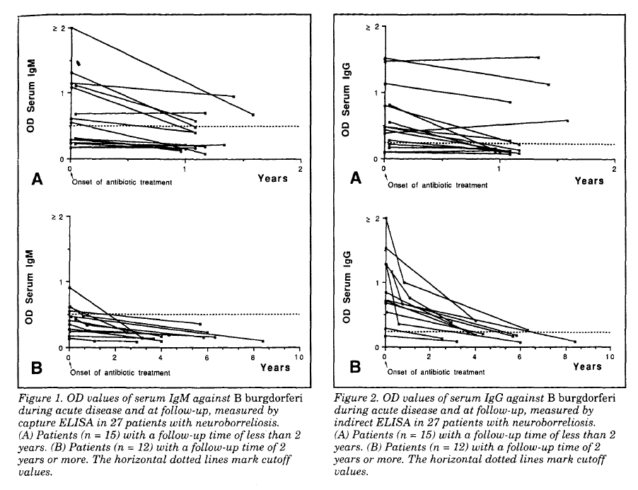
Results. Serum antibody production. The first serum sample from two of 27 patients was negative for both IgM and IgG antibodies against B burgdorferi in ELISA. Both these two seronegative patients had had neurologic symptoms for 2 weeks. Twelve patients had positive serum IgM (figure 1, A and B), and 22 patients had positive serum IgG OD values against B burgdorferi in their first serum sample (figure 2, A and B). Three patients with only positive IgM ELISA had a duration of neurologic symptoms for 2 to 4 weeks (median, 2 weeks). Nine patients who were positive for both IgM and IgG had a disease duration between 3 weeks and 8 months (median, 5 weeks). The remaining 13 patients, who were positive in only IgG ELISA, had a duration of neurologic symptoms from 3 weeks to 2 years (median, 2 months and 3 weeks).
   The kinetics of the IgM and IgG responses in serum after treatment of the 27 patients are shown in figures 1, A and B and 2, A and B. Among 15 patients with a follow-up time of less than 2 years (11.5 months to 1 year and 7 months; median, 13 months), seven patients were still seropositive (figures IA and 2A). Two of the seven patients had only positive IgM measurements two patients were positive for both IgM and IgG, and three patients had only positive IgG OD values against B burgdorferi in serum. Among these seven still-seropositive patients at follow-up, only two had a significant decline in IgM ELISA (figure 1A), whereas IgM OD values in two patients and IgG OD values in the five patients who were still IgG-positive showed no significant changes (figures 1A and 2A).
   Among 12 patients with a follow-up time of 2 years or more (24 years to 8 years and 5 months; median, 4 years), none had positive IgM OD values in serum (figure 1B), whereas three patients were still positive in serum IgG (figure 2B). All three seropositive patients had a significant decline of OD values (figure 2B).
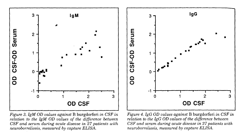
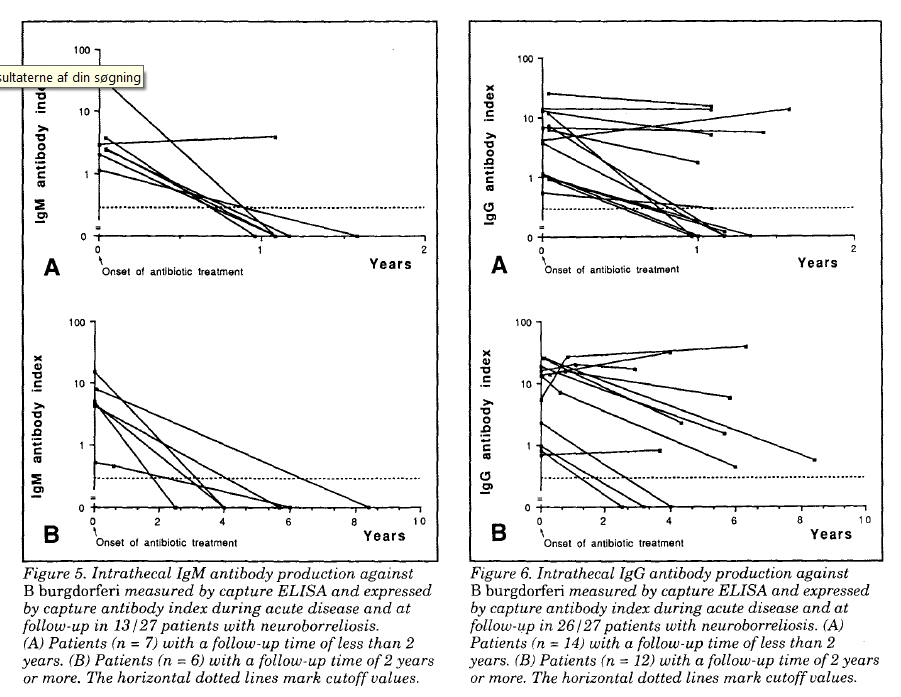
   Intrathecal antibody production. According to inclusion criteria, all patients had signs of intrathecally produced IgM and/or IgG antibodies against B burgdorferi in the first pair of serum/CSF samples (figures 3 and 4). Thirteen of 27 (48%) patients had a positive IgM antibody index (figure 5, A and B) and 26/27 (96%) had a positive IgG antibody index (figure 6, A and B). One patient had only a positive IgM antibody index, 12 patients had both positive IgM and IgG antibody indices, and 14 patients had only a positive IgG antibody index. The patient with only a positive IgM antibody index had a duration of neurologic symptoms for 2 weeks, whereas the 12 patients with both positive IgM and IgG antibody indices had had neurologic symptoms for 2 weeks to 8 months (median duration, 1 month). Fourteen patients with only a positive IgG antibody index had a duration of neurologic symptoms between 3 weeks and 2 years (median duration, 4 months and 1 week). The kinetics of IgM and IgG antibody indices are shown in figures 5, A and B, and 6, A and B.
   At follow-up, only one of 13 patients (8%) with an initial intrathecal IgM production against B burgdorferi had a positive IgM antibody index (figure 5A). This patient had a follow-up time of 13 months.  Fifteen of 26 patients (58%) with specific intrathecal IgG synthesis during acute disease had a positive IgG antibody index at follow-up. Six of these 15 patients had a follow-up time of less than 2 years (figure 6A), and the remaining nine patients had been followed for 2 years or more (figure 6B).
   Serum antibodies and intrathecal antibody production. During acute disease, five patients had signs of specific intrathecal IgM production against B burgdorferi and negative IgM in serum, and four patients were positive in serum IgM without signs of intrathecally produced IgM. Four patients with negative serum IgG OD values had intrathecal IgG synthesis against B burgdorferi.
   At follow-up, signs of intrathecal antibody production against B burgdorferi had disappeared in 11 of the 27 patients. Two of these 11 patients were still seropositive: one had a positive IgM OD value and the other a positive IgG OD value.
The only patient with persistent intrathecal IgM production at follow-up was seronegative in both IgM and IgG ELISA.

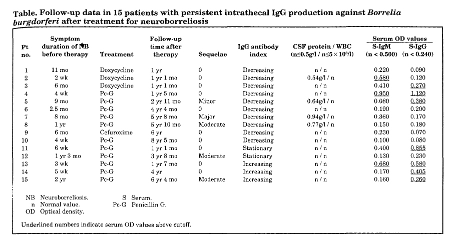
Among the 15 patients with persistent intrathecal IgG synthesis, eight patients remained seropositive at follow-up. One patient still had only a positive IgM OD value, two patients had both positive IgM and IgG measurements, and five patients had only positive serum IgG OD values against B burgdorferi (table).
   Clinical follow-up findings in patients without persistent intrathecal antibody production against B burgdorferi. Among the 11 patients without persistent specific intrathecal antibody synthesis, nine patients, all with early neuroborreliosis during acute disease, had a complete clinical recovery. One patient with early neuroborreliosis had minor sequelae; another, with late neuroborreliosis, had major sequelae. The patient with major sequelae had symptoms of ataxia, visual impairment, and a slight pleocytosis at follow-up. Compared with pretreatment 14 months earlier, she had improved.
   Follow-up data in patients with persistent intrathecal antibody production against B burgdorferi. The only patient with persistent intrathecal IgM production against B burgdorferi 13 months after treatment with doxycycline had a negative IgG antibody index, a normal protein level and normal cell count in CSF, and a complete clinical recovery. She had had symptoms of neuroborreliosis for 2 weeks before treatment. All 15 patients with positive IgG antibody indices at follow-up had improved compared with pretreatment, and none showed progression or signs of disease activity. Among these 15 patients with persistent intrathecal IgG production against B burgdorferi, nine patients with early and one patient with late neuroborreliosis had a complete clinical recovery. Five patients, one with early and four with late neuroborreliosis, had sequelae at follow-up. The individual clinical and laboratory findings at follow-up in these 15 patients are listed in the table.
   Controls. Sera from three controls showed positive IgM OD values against B burgdorferi; in two these controls, IgM measurements were slightly elevated, at OD 0.620 and OD 0.650. One of them suffered from tick-borne viral encephalitis and the other from echovirus meningitis.
[? => Can echovirus perhaps be transmitted by ticks too?]
The third control patient had a moderately elevated IgM in serum, OD 1.240. This patient had meningococcal meningitis.  Two other controls had positive serum IgG OD values against B burgdorferi, one with a moderate elevation, OD 0.970, and the other with high serum IgG measurement, OD 2.550. Both these patients had herpes zoster meningoencephalitis. None of the controls had signs intrathecal IgM or IgG antibody synthesis against B burgdorferi.

Discussion. The demonstration of a specific intrathecal immune response against B burgdorferi together with a lymphocytic pleocytosis is the best laboratory evidence of active neuroborreliosis. The capture technique20 offers some advantages in comparison with the conventional method of analyzing serum and CSF by indirect ELISA.18,22-24
23. Hofstad H, Matre R, Nyland H, Ulvestad E. Bannwarth’s syndrome: serum and CSF IgG antibodies against Borrelia burgdorferi examined by ELISA. Acta Neurol Scand 1987;75:37-45. PMID: 3577666
24. Wilske B, Schierz G, Preac-Mursic V, et al. intrathecal production of specific antibodies against Borrelia burgdorferi in patients with lymphocytic meningoradiculitis (Bannwarth's syndrome). J Infect Dis 1986;153:304-314. PMID: 3944483
With the capture ELISA, the relative amount of specific to unspecific antibodies in CSF can be directly compared with the corresponding ratio in serum, thus identifying the presence of a specific intrathecal antibody synthesis without correction for the leakage over a damaged blood-CSF barrier. The use of purifďed B burgdorferi flagella as antigen has earlier been shown to be of advantage in assays for diagnosis of neuroborreliosis.16,22,24 The importance of measuring a specfic intrathecal antibody response is demonstrated by the fact that two patients with early neuroborreliosis were seronegative and by the results obtained among the control patients in the present study. Whereas five of the 37 control patients were seropositive, none had signs of intrathecally produced antibodies against B burgdorferi. Positive serum OD values in the controls might be explained by either unspecific reactions or previous subclinical or clinical infection with B burgdorferi.
   The measurement of the intrathecal antibody response is more complex in comparison with the analysis of the serum antibody response. The index value is influenced not only by the proportion of specific antibodies in CSF and serum but also by the blood-CSF-barrier permeability. The kinetics of the antibody synthesis in serum and CSF may differ, and a more rapid decline of the specific antibody production in serum compared with the production in CSF will give an increasing index. Furthermore, in patients with a severely damaged blood-CSF barrier during active disease, the relative amount of specific antibodies synthesized in CSF will increase when the blood-CSF barrier normalizes, due to the disappearance of a large proportion of specific and unspecific serum antibodies from CSF. This may cause an increasing antibody index after treatment despite stationary or even decreasing specific antibody synthesis in both serum and CSF. Thus, the comparison of the numeric value of the specifIc IgG antibody index in consecutive samples in individual patients with neuroborreliosis is difficult, and of less importance for treatment evaluation.
   Since follow-up samples were most often drawn only once in each patient, it is not possible to analyze the true kinetics of the intrathecally produced antibodies. However, in two of four patients in the study from whom consecutive serum/CSF samples were available, there was an initial rise of IgG antibody index compared with pretreatment level before a final decline. This is in agreement with the results of Hansen and Lebech.20
   More than one-half of the patients in the present study had persistent intrathecal IgG production against B burgdorferi at follow-up. Generally, there was a tendency toward decreasing values of the capture antibody indices over time. Consequently, the frequency of patients with persistent specific intrathecal IgG synthesis depends on the follow-up time. Compared with persistent seropositivity, only three of the 12 patients with a follow-up time of 2 years or more still had positive serum IgG OD values, whereas nine of these 12 patients had ongoing intrathecal IgG antibody synthesis. Thus, the frequency of persistent intrathecal antibody production seems to be higher than the persistence of seropositivity over time after neuroborreliosis.
   A few previous publications11,16,20,25
25. Martin R, Martens U, Sticht-Groh V, Dörries R. Krüger H. Persistent intrathecal secretion of oligoclonal, Borrelia burgdorferi-specific IgG in chronic meningoradiculitis. J Neurol 1988;235:229-233. PMID: 3373242
have shown intrathecal immunoglobulin production against B burgdorferi for ˝ to 1 year after antibiotic treatment of neuroborreliosis. Intrathecal antibody response against B burgdorferi has been present months to several years after spontaneous recovery from neuroborreliosis, with or without sequelae.11,19,24,26
26. Henriksson A-M, Link H, Cruz M, Stiernstedt G. Immunoglobulin abnormalities in cerebrospinal fluid and blood over the course of lymphocytic meningoradiculitis (Bannwarth’s syndrorne). Ann Neurol 1986;20:337-345. PMID: 3532931
The results of the present study show that, occasionally, specific intrathecal antibody production may be demonstrated for almost 10 years after successful antibiotic treatment of neuroborre1iosis. No earlier publication has shown persistent intrathecal antibody synthesis against B burgdorferi for such a long time after therapy. Theoretically, this finding may represent a latent infection.11 However, since all the patients had been treated and showed no signs of clinical progression or relapse during the several years of follow-up, an anamnestic remaining antibody response is a more probable explanation. [MK: I'd like to ask these patients personally today, if they are still 100% okay.]
   In all patients but one, there was a good correlation between the clinical recovery from neuroborreliosis and the disappearance of intrathecally produced IgM antibodies against B burgdorferi. The disappearance of intrathecal IgM antibody production may be a spontaneous event, but is more likely explained by the elimination of antigen due to antibiotic treatment. The finding of persistent specific intrathecal IgM synthesis more than 1 year after treatment in one patient with a complete clinical recovery is somewhat surprising, and so far we have no explanation.
   There was a tendency toward more patients with sequelae (five of seven) having persistent intrathecal IgG antibody production against B burgdorferi than patients without sequelae (10/20). One possible explanation is persistence of Borrelia spirochetes in CNS in patients with sequelae. However, a more likely theory, in our opinion, is that the majority of patients with sequelae and intrathecal IgG synthesis against B burgdorferi at follow-up had late or chronic neuroborreliosis (four of five), with disease durations of several months or even years before antibiotic treatment. Previous studies have shown that the antibody synthesis against B burgdorferi in CSF increases with time, resulting in higher CSF antibody titers in patients with a long disease duration.18 It seems probable that such a 1ong1asting antibody response might persist for a long time after the antigenic stimulation has disappeared after antibiotic treatment.
   The results of the present study show that intrathecally synthesized antibodies against B burgdorferi, mainly IgG, may be present for several years after successful antibiotic treatment of neuroborreliosis. Consequently, the finding of intrathecal IgM and IgG production against B burgdorferi must always be evaluated in relation to clinical data and signs of inflammation in CSF, and does not necessarily indicate active neuroborreliosis nor the need of further antibiotic treatment.

Comment by MK. These authors forget to take into consideration the following::
  1. In a currently active Borrelia infection with surplus of ANTIGEN compared to ANTIBODY, it can happen that all antibodies get quickly trapped in immune-complexes, so NO ANTIBODIES ARE FREE and able to bind til the TEST FLAGELLA, which then measures nothing, i.e false seronegative test outcome 
  2. Hardin et al. 1979 (PDF) found amount of circulating immune complexes in Lyme arthritis that was high in early infection, highest in the sickest, low/absent during remission and fluctuated with recurrent clinical activity
    Schutzer et al. 1990 (PMID: 1967770) (which Hansen ref. in another of his publication in 1994, thus he is aware of the paper!) demonstrated Borrelia burgdorferi specific circulating immunecomplexes in 10 seronegative active Lyme disease cases, later confirmed by many others; references and danish comment by MK in http://kroun.ulmarweb.dk/Borrelia-IC.html
  3. 1985 PMID: 3542350 mention in abstract that "Antigenic variation between isolates may determine the differences in clinical expression observed between cases in North America and Europe." - antigenic variation was already wellknown for closely related relapsing fever Borreliae.
  4. 1992 Baranton et al. (PDF) publiced finding 3 genetically different Borrelia strains associated with different clinical pictures, Borrelia burgdorferi sensu stricto (arthritis, USA + EU), Borrelia garinii ("typical neuroborreliosis", lymphocytic meningoradiculits AKA Bannwarth syndrome), Borrelia afzelii (VS461; since old times associated with chronic multi-organic manifestations, incl. joint and CNS and even bone marrow inflammation; arthritis plus ACA has been described in EU since 1920'ies, see some older refs. in http://lymerick.net/Bb-history.pdf .. 
  5. 1992 Picken (PDF) published on variations in the flagellin gene, which implicate possible resulting differences in the flagella antigen among different Borrelia strains, so flagellin anitbodies raised by mammal host to other Borrelia strains with differing flagella antigens, may not be able to bind til the Borrelia afzelii DK1 flagellin based antigen!? - hence could result in false negative test results.
  6. Hansen & Lebech in J Clin Microbiol. 1992 Jul;30(7):1646-53. PDF page 1652: "B. burgdorferi antigen has never been detected in the CSF of patients with neuroborreliosis." and "However, clinical experience has never revealed the occurrence of relapses in patients with neuroborreliosis who were appropriately treated (22, 29)." - these postulates had both been disproved by culture verified published cases see http://lymerick.net/persistent-borreliosis.htm
    Preac Mursic V, Weber K, Pfister HW, Wilske B, Gross B, Baumann A, Prokop J. Survival of Borrelia burgdorferi in antibiotically treated patients with Lyme borreliosis. Infection 1989 Nov-Dec; 17(6): 355-9 PMID: 2613324
    1. 5y boy. July 1985 EM. Aug 1985 Lymphocytic meningitis. Seropositive for IgG and IgM, no antibodies in CSF. Penicillin V orally 100000 u/kg/d, 14d. Spinal-fluid showed fewer cells. September 1985 facial palsy, again pleocytosis in CSF. Doxyc. orally 2mg/kg, 10d. Gradually CFS normalized. April 1986 relapse, Bb was isolated from CSF after 4 weeks in BSK-medium. Penicillin 200000 u/kg, 22d. August 1986 relapse/reinfection with EM and painful meningoradiculitis, Bb antibodies now negative in CFS and serum. Culture not done!
    2. 49y man. EM, typical signs of LMR-Bannwarth S developed 7 weeks later. Pleocytosis and elevated protein in CSF. Both Borrelia IgM and IgG positive in serum. Penicillin i.v. 20 MU/d, 10d. Four days after therapy normal examination and no complaints, CSF declining parameters, positive Borrelia-index. Three months later CSF normal, Borrelia-index now negative, but Bb was cultured from CSF!
    3. 26y woman. Headache, radicular pain. Normal neurological exam. Multiple horseflie bites. CSF pleocytosis and elevated protein. Negative Borrelia-ELISA in CSF and serum. Ceftriaxone i.v. 2g/d , 10d. Improved. 7.5 month later recurrent episodes of radicular pain, headache, arthralgia, fever. Normal neurological exam. Negative serology. Normal CSF. Bb cultured from CFS after 6 weeks in MKP-medium. Cefotaxime 3 x 2g/d i.v., 14d.
    .... 3 skin isolates in post treatment relapses also descrihed ...

    Many more articles on culture, microscopy or PCR proven cases have been added since, and particularly two studies are worth mentioning in this context, because they both used DAKO EIA (Hansen's Borrelia flagella based antibody test):
    Oksi J Clin Microbiol 1995 (PDF): 41 late (> 3 mdr.) borreliosis patients with either positive culture (12) and/or positive PCR (39); DAKO FL-ELISA missed 6 of 12 culture verified cases or 24/41 (58%) of culture and/or PCR positive cases.
    Strle Clin Inf Dis 2006 (PDF): 33 in CSF culture-verified neuroborreliosis cases; 23 Borrelia garinii (which fitted the "typical" picture of neuroborreliosis, clinically and in laboratory measures), however 10 patients with Borrelia afzelii neuroborreliosis, differed clinically only significantly in one symptom (painful radiculitis), only 3 had been sick < 3 months; 7/10 (70%) DAKO EIA CSF/serum index negative, despite duration of symptoms from 6 months to 7 years!     
Conclusively:

Several WARNING SIGNS AGAINST OVER-GENERALISATION had been published, some even before, others after these authors - in Brain 1992 - expressed their BELIEF and claims, that SERUM IgG measured by their flagella antibody test will always become positive at latest 3 months after symptom debut, and that all neuroborreliosis patients will develop positive CSF/SERUM index measured with their flagella antibody test, at latest within 8 weeks after symptom debut
- statements since adopted by Statens Serum Institut (SSI), the danish national microbiological reference laboratory link (danish), which continue to maintain these postulates still, despite the published evidence AGAINST!
First, Hansen and Lebech have self reported a few cases (also here!), that to their surprise, did not develop positive Borrelia flagella-IgG in their serum > 3 months (cases were usually spinal Borrelia Index positive, in this paper only IgM-index positive)!
Secondly, published works listed in http://lymerick.net/persistent-borreliosis.htm (available for all to see on the Internet since 2003), for instance the paper published by Oksi et al.. in J Clin Microbiol 1995, in which a total of 24/41 (58%) with DAKO test measured SERONEGATIVE, BUT CULTURE or PCR PROVEN LATE (all sick > 3 months) BORRELIA CASES AGAINST Hansen et al's OPINION! - plus further evidence against has piled up since ...
POSITIVE CULTURE IS THE VERY BEST POSSIBLE MICROBIOLOGICAL EVIDENCE ONE CAN GET FOR CURRENT BACTERIA INFECTION, and culture is often called the GOLD STANDARD MICROBIOLOGICAL DIAGNOSTIC METHOD! 

Looking deeper into the references which Hansen and Lebech uses to back up their statements (sometimes they do not reference any published works), the above underlined claims seem not to be SCIENTIFICALLY EVIDENCE BASED!
First, their main research studies on the flagella based serology test were done in clinically highly selected patient group, all must express "typical neuroborreliosis" AFA lymphocytic meningoradiculitis AKA Bannwarth S, which has (since Baranton 1992) been found especially associated with infection with a particular strain of Borrelia burgdorferi sensu lato*, i.e. Borrelia garinii that seems to cause more pronouced host immune and CSF reactions, compared to other strains of Borreliae during infection of the nervous system or without infection of the nerve system.
Next, Hansen and Lebech have (paradoxically it seems in hindsight) selected a different strain of Borrelia burgdorferi sensu lato (B. afzelii) DK1 for their flagella antibody test, which was/is mainly intended for diagnosing "typical" neuroborreliosis(!); it appears from their statements that they have believed flagella antigen to be highly conserved ~ exactly the same in all borrelia strains, at least until Picken 1992 (which Hansen and Lebech references in 1994!) showed the flagella gene variation among borrelia strains; but Hansen and Lebech have not taken the logical consequence of this knowledge, which would have been to shift test antigen to Borrelia garinii flagella, after the genetic differences among different borrelia strains became revealed ...
Since, they have failed to modify the claims, that SERUM IgG, measured by their flagella antibody test method, will always become positive at latest 3 months after symptom debut, and that all neuroborreliosis patients will develop positive CSF/SERUM index , again measured with their flagella antibody CSF index test, at latest within 8 weeks after symptom debut
- statements that were adopted by Statens Serum Institut (SSI), the danish national microbiological reference laboratory link (danish), which continue to maintain these postulates still (checked May 2010), despite scientific evidence against and the resulting misuse by danish doctors against have actually been presented to SSI by me, who also pointed ouf the LACK OF DECLARATION OF HANSENS CONFLICT OF INTEREST, over one year ago! 
SSI / Hansen et al. have failed to do a similar controlled study in culture or PCR proven DANISH LATE BORRELIOSIS CASES, as Oksi, i.e. has failed to prove with same strengt as the opposing science, that their claims do hold; futhermore I have suggested such a study to SSI and also suggested co-work about such a study to the local university hospital microbiology department, since I have access to the ideal supected chronic Borreliosis patients for such a study in the patient society; NOT INTERESTED was the answer from the leading microbiologist (2008)!
These authors / SSIs statements / "expert opinions" may possibly hold up for and be somewhat justified by Hansens et al's findings done in their specially selected group "typical" neuroborreliosis - most probably caused by Borrelia garinii - cases, that is if these authors have NOT intentionally excluded consecutively enrolled patients clinically suspect of having neuroborreliosis, by the fact that some of these patients did NOT become seropositive within their time limit ... which they very possibly might have done, since a positive spinal index (within a month after symptom debut?) has often been a strictly required inclusion criteria in several of their studies! - thus they could perhaps have excluded some serum and/or CSF antibody flagella antibody negative, bu truly Borrelia infected with other strains of Borrelia,  from participating in their serology studies, which may then have resulted in skewed results, a wrong impression!?
- thus it seems Hansen and Lebech have made a circular argument / conclusion (Wiki)! 
 
The danish authors - Hansen and Lebech - were co-authors on a danish consensus report / officially recommended Borrelia guidelines from 2006 (link 2010 version corrected), which fails to discuss the above mentioned caveats.
The working group has firmly advocated using ONLY DAKO/OXOID Borrelia flagella antibody test  for the diagnosis of Borreliosis in Denmark, the very same test which Hansen has just (May 2010) been forced by the press (INFORMATION.DK) to admit that he co-owns and earns money on by sale, and which he has "forgotten" to declare his CONFLICT OF INTEREST - that is, not only in the Borrelia guidelines report from 2006 - but also in other published papers, which he has co-authored, since the rules have for a long time dictated that such a declaration of eventual conflicts of interest is necessary for all authors of scientific papers to send in!
Moreover, in the 2006 danish Borrelia guidelines also fails to mention / discuss / reference Oksi's paper, which PROVES Hansen and Lebech's IGNORANCE of certain counter proof!
The guideline report also failed to address the important implication of antigen trapping of some/all formed antibodies in immune complexes, during times of ACTIVE BORRELIOSIS (early or late).
Besides the authors have twisted some statements in the guidelines report, so it fits their viewpoint [chronic seronegative Lyme borreliosis does not exist, stated by Hansen in danish article in MPL 1994] a little better, than what is actually accounted for in the source text behind the reference; twists that will only be noticed by readers very well into the Borrelia literature already and/or those who decide to check what is really stated in the referenced source text!

In this paper  - Neurology. 1993;43:169-175 - Hansen and Lebech fails to mention the very important 1.5 time increase in IgG cut-off value, which it seems is not being backed up by scientifically peer-review published literature; at least I have not been able to find the cut-off value validation that find OD of 0.240 for IgG, in any of the sources to the sources, they have chosen to reference as backup for the increase; most of the older papers referenced use OD 0.160 as the 98% (some 95%) percentile cutoff value for IgG! - therefore the increase in IgG cut-off, does not seem to be SCIENTIFICALLY PROVEN / IS NOT EVIDENCE BASED / IS NOT JUSTIFIED?!  
WHY IS THIS INCREASE In IgG CUT-OFF NOT EXPLAINED / NOT MENTIONED / NOT DISCUSSED AT ALL IN THIS  FIRST PUBLISHED PAPER USING THE HIGHER IgG CUT-OFF VALUE ?!
- it "smells" of a possible hidden agenda ...
We can wonder why this increase in IgG cut-off came shortly after these authors statement, that neuroborreliosis may be ruled out - authors intention is that no further investigation of the patient for Borrelia infection is needed - if/when the patients does not have  apositive Borrelia IgG in serum, measured by their flagella antibody test, at latest 3 months after symptom debut
- a postulate that is very problematic, especially for the truly Borrelia infected patients with hampered immune function, for instance those having co-infection(s) with immune cell infecting microorganisms, and/or have other immune depressing co-factors involved in their disease process
- because after the increase in cut-off it has become 1.5 times harder for the patient to get a positive SERUM Borrelia IgG result?! 

*) In Europe until now 6 strains within Borrelia burdorferi sensu lato complex has been isolated by culture or detected by PCR in European HUMAN cases, besides Borrelia burgdorferi sensu stricto, Borrelia garinii, Borrelia afzelii, also:
Borrelia valaisiana: 2004 EID; 10 year neuroborreliosis, pos. PCR in CSF, seronegative. Thus is NOT ref. in the danish guidelines report, which instead states, that human infection with Borrelia valaisiana has not yet been proven!
Borrelia lusitaniae:  2005 PMID: 16053200, PDF; 10 year old skin affection, culture pos., seronegative;  in the PubMed abstract is stated that the pt. never had an EM, but this wrongly was told to be EM in the danish consensus report from 2006!
Borrelia spielmanii: 2005 EID, culture skin EM, seronegative, lost to follow up; there are over 100 known strains of Borrelia detected in ticks that must somehow maintain a tick-mammal lifecycle, which has not YET been proven to be able to infect and be pathogenic for humans; the perspective is that there may be plenty of human Borrelia infection falsely outruled by negative serology status, that we won't find out about if they are human infective and pathogenic, until we have tests available, that are able to detect all the various borrelia strains; serology is highly problematic and will always be problematic, because of Borreliae ability to undergo rapid antigenic variation, and ability to alter into more resistant alternative forms (100 years pictorial, Burgdorfer 1999 keynote lecture) that may let Borrelia survice any adverse conditions, including even long term antibiotic treatment! - later, when Borrelia growth is no longer suppressed sufficiently, some hidden forms may revert back to spirochete form and cause clinical and with direct test methods confirmed relapse of Borrelia ACTIVITY; Borrelia uses many different tricks to evade mammal host immune attack and Borrelia may even cause (Borrelia specific?) immune depression in LATE infection, for instance indicated by Dorward 1997 PDF - if Borrelia get enough time (late long tern untreated Borrelia infection) and have success to invade and kill all the host memory cells that are directed against Borrelia's surface antigens, this would give a probable explanation, why so many patients, that initally in the infection responded normally with production of Borrelia antibodies, but which after a while, stop producing these antibodies, despite they have clinical relapse and by direct methods verifiable persistent and active Borrelia infection!?

WE NEED ACCESS TO DIRECT DETECTION METHODS FOR LATE DIAGNOSED / CLINICALLY SUSPECTED RELAPSING LYME BORRELIA CASES; such tests are not currently supplied by any danish microbiology laboratory!
POSSIBLY PROPER EVALUATION IS BEST DONE ABROAD - in other EU countries like Germany, which have many more Borrelia infections yearly, therefore - compared to danish doctors - have much more  experience with chronic / relapsing Borrelia cases, than it is possible to get for any doctors here in Denmark, where only 100-200 neuroborreliosis cases yearly are registred officially, of which "only" ~ 10% (10-20 pt.) yearly (the % of possible relapses in Denmark was deducted from Hansen and Lebech, Brain 1992; others researchers find up to 20% relapses after recommended treatment) may relapse / have persistent Borrelia infection  ... because of much larger number of patients in need, these laboratories can afford to run an array of other supplementing tests, including PCR, also for other (tickborne) co-infections, which those patients that relapse from Borreliosis must also be investigated thoroughly for, of course!
Acknowledgment
We thank Dakopatts, Copenhagen, Denmark, for supplying us with the Dako Lyme Borreliosis ELISA kit K 416.


Table:
8423881-table.jpg
Figures:
8423881-fig1-2.jpg
8423881-fig3-4.jpg

8423881-fig5-6.jpg