Morphologic
characteristics of certain cultured strains of
oral spirochetes and Treponema pallidum
as revealed by the electron microscope.
Hampp EG, Scott DB, Wyckoff RWG
J Bacteriol 1948;56:755-69
48_02 Morphologic char spirochet & TP EM.tif [Imaging for W95B+]
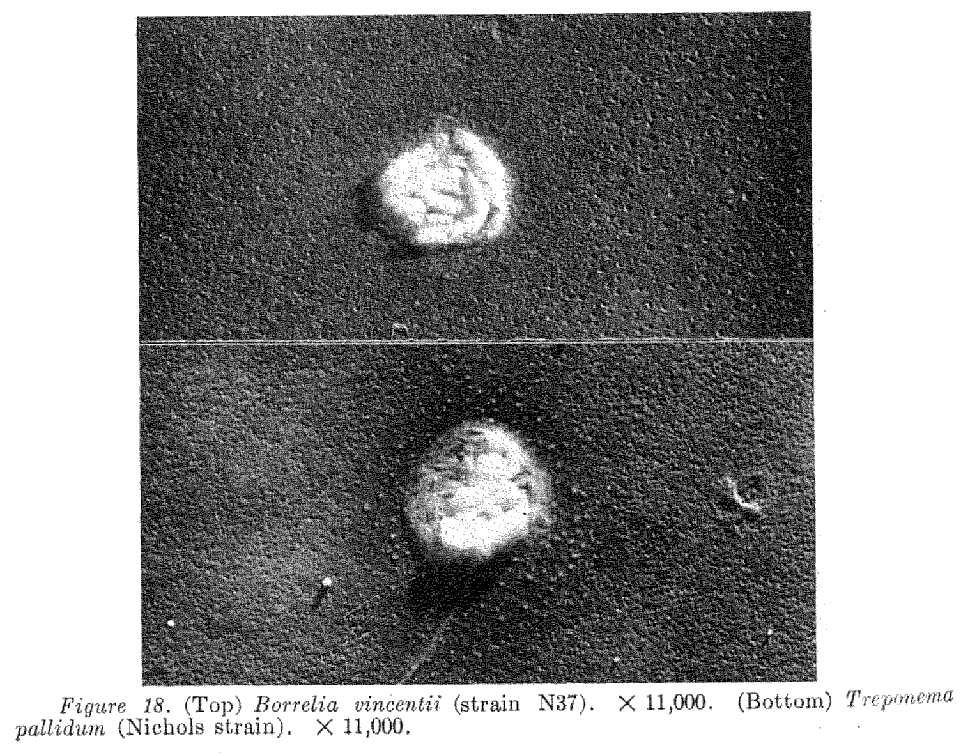
Shadowed
preparations of pure cultures of two strains of the small oral treponemes, four strains of Borrelia vincentii,
and the Nichols and Noguchi cultured strains of Treponema
pallidum have been studied with the electron
microscope. Morphological characteristics, filamentous and flagellar
appendages, and granules of various types have been described and illustrated.
[the granules resembles what Brorson
and Alban call 'cyst', see Spirochetal-cysts.htm ]

Excerpt:
Typical free granules, the end products of granule 'shedding', are shown in
figure 18.
They are roughly circular in
outline and sharply bounded. They consist for the most part of what appear to
be short sections of spirochetes closely packed together. The
contents of these granules are probably responsible for the fine lacelike appearance
and the bright white, highly refractile bodies
described by Hampp (1946) under the dark-field
microscope.
Examples of another type of free granule repeatedly observed are shown in
figures 19 and 20. These granules consist of tangled masses of spirochetes or spirochetal segments.
The significance of granules in the
life history of the spirochetes is unknown but certain investigators have
suggested that they may be germinative units (Balfour,
1911; Noguchi, 1911; Noguchi, 1917; Leishman, 1918; Mudd et al., 1943; Hampp, 1946).
Others are undecided or hesitant in accepting this hypothesis (Fantham, 1916; Akatsu, 1917; Wenyon, 1926; Warthin and Olsen,
1930). Topley and Wilson (1936) have indicated that
they are probably particles of culture medium adhering to the sides of the
spirochetes. The electron micrographs demonstrate that this explanation is
wrong, and that free granules
are definitely a phase in the development of spirochetes. Although
it is not possible to determine from these micrographs that the granules are germinative units their constant rhythmic occurrence in
living cultures suggests this possibility. Further support of this hypothesis is provided by the fact
that cultures up to 31 months old, showing only refractile
granules by dark-field examination have invariably given normal growths on
transfer to fresh medium (Hampp, 1946)
* * *
That
the long time in 'granule' form can explain relapses of spirochetoses
for equally long after the primary infection, has later been supported by:
Oksi et el. Borrelia burgdorferi detected by culture
and PCR in clinical relapse of disseminated Lyme borreliosis.
Ann Med 1999 Jun;31(3):225-32. (full
text online unfortunately no longer available)
where patient no 2 relapsed 130 weeks after first antibiotic treatment, no
suspicion of reinfection in the meantime.
The patient had both IgM and IgG
antibodies at first diagnosis, but was seronegative at time of clinical and PCR-proven relapse of borreliosis
in plasma.
Brorson's and Alban - both describes formation of Borrelia burgdoferi cysts and their transformation back to mobile
spirochetes again:
http://www.ncbi.nlm.nih.gov/entrez/query.fcgi?cmd=search&term=(Brorson+OR+Alban+AND+Borreli*)&dispmax=1000&doptcmdl=abstract
see 1998-Brorson-cyst.htm
for excerpts. Brorson describes herein that one cyst can liberate up to at
least 5 new spirochetes.
The infectivity of the Borrelia garinii cyst
form has been shown by Gruntar et al:
http://www.ncbi.nlm.nih.gov/entrez/query.fcgi?cmd=Retrieve&db=PubMed&list_uids=11478686&dopt=Abstract
Abstract:
Cystic forms (also called spheroplasts or starvation forms) and their ability to
reconvert into normal motile spirochetes have already been demonstrated in the
Borrelia burgdorferi sensu lato complex. The aim of this study
was to determine whether motile B. garinii could
develop from cystic forms, not only in vitro but also in vivo, in
cyst-inoculated mice. The cysts prepared in distilled water were able to
reconvert into normal motile spirochetes at any time during in vitro
experiments, lasting one month, even after freeze-thawing of the cysts. Motile spirochetes were successfully
isolated from 2 out of 15 mice inoculated intraperitoneally
with cystic forms, showing the infectivity of the cysts. The demonstrated capacity of the cysts to reconvert
into motile spirochetes in vivo and their surprising resistance to adverse
environmental conditions should lead to further studies on the role and
function of these forms in Lyme disease.
Marie Kroun, MD
April 2001
Links edited July 2006