[The following is a transcript of article in Ann Inst Pasteur 1918, 32:49-59 /Marie Kroun 09-09-2000]
ANNALES
DE
L'INSTITUT PASTEUR
Mémoire publie à 1'occasion du jubilee de E. Metchnikoff.
A NOTE ON THE "GRANULE-CLUMPS"
FOUND
IN ORNITHODORUS MOUBATA
AND THEIR RELATION TO THE SPIROCHAETES
OF AFRICAN
RELAPSING FEVER (TICK FEVER)
by Colonel Sir WILLIAM B. LEISHMAN
C.
B., M.B., F. R.C. P., F.R.S., LL.D., K.H.P., A.M.S.
In 1908 I obtained a number of Ornithodorus moubata from Nyasaland and from Uganda, with a view to studying the course of events in Tick Fever in experimental animals. After several failures I eventually secured a batch of ticks which proved to be infective and by their bites produced in a monkey. a typical attack of Relapsing Fever, associated with the occurrence of large numbers of Spirochaeta duttoni in the blood. I was then able to infect a number of ticks by allowing them to gorge themselves on this infected monkey and have, since then, been able to maintain the strain of spirochaetes, either by passage from mouse to mouse or through the bites of infected ticks. I was thus in a position to study the fate of spirochaetes ingested by the ticks, as well as such questions as the nature of the hereditary transmission of the infection in the tick, the mechanism of infection by the bite of the tick, and other points. The results of some of this work were published in 1909 and 1910 [1].
The purpose of the present note is to record some further observations which appear to throw fresh light upon the nature of the granule-clumbs, descibed in the above mentioned papers, which, I suggested, were probably derived from spirochaetes and were capable, under certain conditions, of once again developing into spirochaete form. To recall a few of' these earlier observations; it was noted that these granules were almost constantly found in various tissues of Ornithodorus and that the inoculation of tissues containing such granules, but, as far as could be determined no spirochaetes, frequently resulted in the production of spirochætosis in mice. The occurrence of similar granules in the eggs of the fecundated female tick and my almost invariable failure to find spirochaetes in such eggs, even when the mother tick had heen heavily infected shortly before, further suggested to me that it might be in this form that the virus passed to the next generation of ticks.
Some of my experiments have since been repeated by other workers, either with Spirochaeta duttoni and Ornithodorus moubata or with Spirochaeta gallinarum and Argas persicus with contradictory results. Thus, Hindle [2] and Fantham [3] confirmed many of my observations and agreed with most of my conclusions, while strong confirmatory evidence was also obtained from Balfour's work on the Spirochaetosis of Sudanese fowls [4] . On the other hand , the more recent work of Marchoux et Couvy [5], of Kleine et Eckard [6], of Wittrock [7] and of Todd [8] has led these investigations to conclusions differing from mine. They have not been able to find any evidence of a connection between the granule-clumps and the spirochaetes and they are satisfied that the facts of' transmission, whether direct or hereditary, are to be accounted for more simply by the persistence of the spirochaetes, as such, in the ticks and by their transmission in this fornm through the egg to the young tick.
I hope before long to lave an opportunity of putting on record fuller details of my recent work than are possible in this note and of discussing some of the apparent discrepancies between the results of my confreres and those of myself. I shall here only mention briefly some recent observations which have strengthened my personal conviction that some, at all events, of these disputed granule-clumps are derived from spirochaetes and are able to subsequently to develope again into spirochaete form.
In my earlier work the method employed was to infect simultaneously a number of ticks of the same age by permitting them to feed on a heavily infected mouse. The ticks were then divided into two or three batches, which were kept under different conditions of temperature and moisture. Every day, or every 2 or 3 days subsequently, one of each batch of ticks was carefully dissected and a close examination was made of the different organs, tissues and fluids for the presence of spirochaetes and of granules; careful records were kept of the results and sketches made of anything of interest. The microscopical examination was in part carried out on the fresh fluids or on pieces of tissue teased out in saline solution, but chiefly by observation of dried film preparations, fixed and stained by my own modification of Romanowsky's method. In the latter case the chromatin staining was carriod to a high degree of intensity, the films being finally treated with 60 p. 100 Alcohol to dissolve off all traces of deposit of the stain. I note that MM. Marchoux et Couvy are of opinion that the Chromatin method of staining may fail to colour thin or delicate spirochaetes and they recommend as a better method the use of Gentian Violet. I find however that my method demonstrates Treponema pallidum readily in films in which these delicate organisnis are quite untouched by Gentian Violet and, further, it permits of the ready recognition of Spirochaeta duttoni in tissues such as muscle fibres when Gentian Violet fails to disclose them.
Subsequently to the series of observations to which my previous papers referred, the dark-ground method of illumination came into more general use and I have since repeated some of my earlier experiments, utilising this method of exanimation as a control and supplement to the staining method mentioned above, and I have found it of great service, particularly in the determination of motility in the spirochaetes. The appearance of spirochaetes illuminated in this manner is naturally familiar to all workers on the subject and I shall only refer to one or two points in connection with it. In the first place, one is struck by the contrast between the high refractility and homogeneous appearance of young and vigorously motile spirochaetes and the low refractility of many of those which are motionless, distorted in shape and presumably dead. Another point which was studied with interest was the so-called <<granule-shedding>> phenomenon, described in connection with Treponema pallidum and other Spirochaetes by Balfour and O'Farrel [9]. In the case of Spirochæta duttoni in freshly shed blood from an infected mouse, kept under continuous observation in a thermostat, it was sometimes easy to detect highly refractile granules, apparently in the substance of the spirochaete, coursing rapidly from end to end of the rapidly rotating organism, and even to observe the sudden extrusion of one of these granules from such a spirochaete. So far, my observations were in accord with those of Balfour and O'Farrel, but I was surprised to find that, if one continued to keep the particular spirochaete and the particular granule which it had extruded under observation, as was sometimes possible, the granule apparently re-entered the body of the spirochaete and, once more, was seen to travel up and down it from end to end. I have observed this sequence of extrusion and intrusion in the case of one spirochaete of one granule, no less than seven times in succession within 3/4 of an hour and I am inclined to think that the <<granule-shedding>> in this instance was an optical phenomenon and could be explained on the assumption that a grain of blood-dust or haemoconia had become involved in the vortex currents produced by the rapidly rotating spirochaete and that it was, in this manner, made to travel up and down the body of the spirochaete, accordingly as as the latter rotated first in one direction and then in the opposite, as is their habit. It is of course quite possible that the granules I observed were not of the same nature as those described by Balfour and O'Farrell, but my impression is that they were the same and that the phenomenon may have a physical rather than a vital explanation.
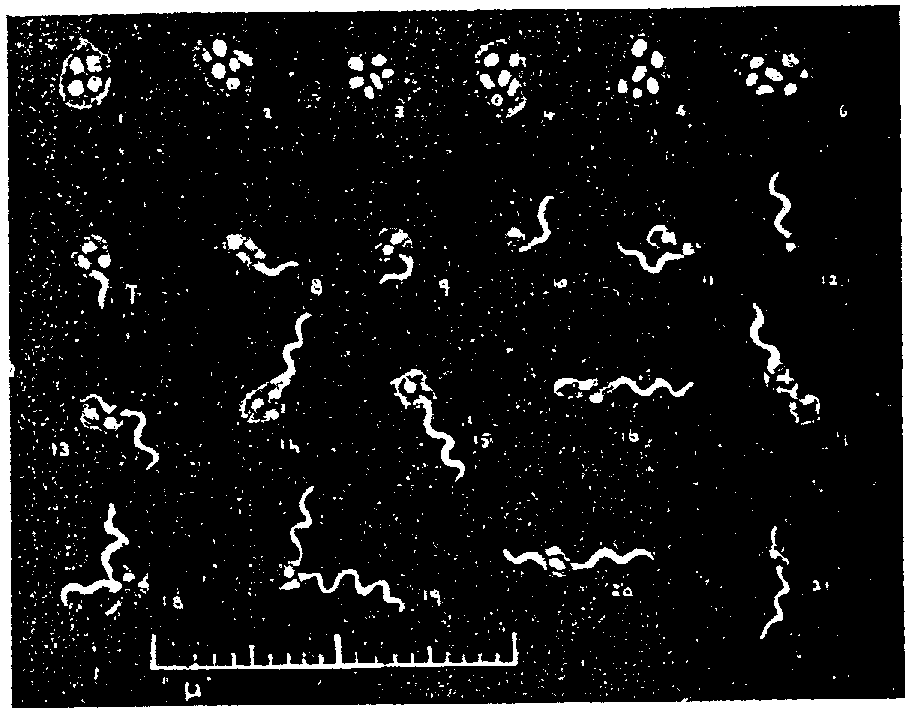
Special attention was paid to the appearance of the granul-clumps under dark-ground illumination. These were most readily observed in crushed portions of a Malpighian tubule or of an ovary suspended in a drop of saline solution. At first it was by no means easy to be certain of the identity of any particular group of granules seen against the dark background, but it soon became possible to recognise them by attention to certain points. In the first place, the size, number and form of the granules composing a clump were more or less uniform; secondly, the brightness and yellowish-white colour of the light refracted by the granules was more or less characteristic and, thirdly, the granules were seen to be embedded in a well defined but faintly refractile matrix, an appearance only occasionally noted in stained specimens. This last point appears to me of some significance as suggesting that there is some vital connection between the different portions of the clump, in opposition to the view that the granules are merely held in apposition by physical attraction. The appearance of the clumps is illustrated in fig. 1, no 4-6.

Figure I.
No 1-6. - << Granule-clumps>> from the tissues of Ornithodorus moubata.
No. 7-10 - Early stage in the extrusion of Spirochaetes from granule-clumps.
No. 11-21. - More advanced stages of extrusion of Spirochaetes from granule-clumps.
In this connection I may mention a point of some technical utility which I have noted in the course of these observations and which is apparently unknown to other workers. It is usually assumed that the use of the dark-ground method of illumination is linited to moist or fluid preparations. I find, however, that it is quite possible to examine a dried and stained film, for example, a film prepared from some organ or tissue of a tick and stained by Romanowsky, to note from this all that can be learned as to the detailed structure of some cell or parasite and then, leaving the specimen in position on the stage of the microscope, to change the illuminating system for the dark-ground method. It will be found that the resulting picture differs very slightly from that which would have been given by the same structure if it had been examined, unstained in a fluid medium. The refractility of the different elements appears little altered and the colours produced by the staining have univ only a slight modifying influence on the white reflex of light refracted from the object. By this procedure it was possible to deternime the staining reaction of a particular granule or granule-clump and then to investigate its refracting power by dark-ground illumination. This method I have found of service in the present investigation and it ought also to be useful in other directions.
In general, this later series of experiments has been confirmatory of my earlier ones but, in several instances, I have found it necessary to revise my former conclusions in the light of further experience. It would not, however, be possible, within the necessary limits of this communication, to deal satisfactorily with the whole of the work; so I shall confine myself to a few points, reserving the fuller account for another occasion.
I find that the spirochaetes, after ingestion by the tick, retain their motility for several days, the period depending chiefly on the temperature at which the ticks have been kept. Subsequently, they lose their motility and tend to agglomerate into great tangles or masses which, when stained, are seen to consist of spirochaetes whose chromatin is fragmented into deeply staining segments or granules. Such spirochaetes I believe to be dead and I think it probable that the segmented chromatin may persist in the form of rods or grains and may reach the Malpighian tubules and other tissues of the tick, accounting in this way for some, perhaps for the majority or the chromatin granules found in such tissues.
Other spirochaetes, however, appear to behave in a different manner and show the curious appearance of a lateral or, more rarely, a terminal protrusion or ''bud''. This lateral bud was described by me in my earlier papers and has also been observed by many others, both in connection with Spirochaete duttoni and other spirochaetes. I am now inclined to attribute to it an important role in the life of the organism, as I believe it to be the origin of the granule clump which may subsequently develope into another spirochaete. I have, for instance, observed a spirochaete which had one of these large lateral buds attached to it, in active rotatory movement for a period of half an hour; the bud contained 3 or 4 highly refractile granules and there could be no possible doubt that the structure was attached to and part of the living organism. At the end of the half hour separation occurred between the bud and the spirochaete, the latter continuing to rotate and bend for another ten minutes, but the separated bud was motionless and corresponded in every particular to the isolated granule clumps to which I have so frequently alluded.
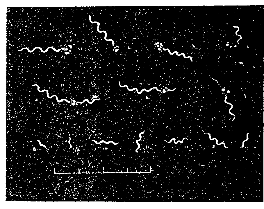
Still later, as one continues to follow the course of events in the batch of ticks, there occurs a period of a few days during which either no spirochaetes at all can be detected or only very rare ones which are seldom motile. Next, and most prominently in the case of ticks which have been kept at comparatively high temperatures (I have experimented with batches kept at 24º, 27º, 30º, 32º, 34º, and 37º C.) there appears a sudden re-invasion of the tissues with numerous and vigorously motile spirochaetes. In some instances I have been able to observe that a large proportion of the spirochaetes which re-appear in this sudden and wholesale fashion are very different in size and general appearance from those which were originally ingested by the tick and which retained their morphological characteristics unimpaired as long as they retained their motility. These ''young forms", as I may call them, are very much shorter than the others, the smallest being from 3 to 4 µ in length and showing only 2 or 3 curves. They are homogeneous and highly refractile and are usually very motile; when stained they take the colour evenly and deeply. Examples of these forms are drawn in figure II. no 8-14.

Figure II.
No 1-7. - Longer forms of Spirochaetes, in association with granule-clumps.
No 8-14. - Young Spirochaetes
The following day, or, days, typical large forms are seen in abundance, as well as young forms and others intermediate between the two.
In the course or several such experiments I have seen this second crop of spirochaetes disappear more or less completely and, six or seven days later, there followed a repetition of the events I have briefly described above. In other words, regular relapses appear to take place in the body of the tick, as regards the appearance and disappearance of the spirochaetes, just as in the case of the warm-blooded host.
In stained specimens of these young forms of spirochaetes, both in the earlier and in the recent series of experiments. some have been seen which were in apparent connection with a chromatin granule or clump of granules, suggesting to my mind a possible development of such a spirochaete from the granule. In the recent work with dark-ground illumination careful watch has naturally been kept for the occurrence of such forms and in four ticks, two of them members of the same batch and the other members of different batches and different experiments, I have observed the forms drawn in fig. I, no 7-21 and in fig. II, no 1-7.
It will be seen from these sketches that many forms have been observed, from the apparent extrusion of a short, highly refractile tail from the granule clump (fig. I, no 7-10) up to typical spirochaete forms showing numerous and regular curves. The forms figured were, with hardly an exception, in active movement at the time of their observation, the longer ones showing the characteristic rotatory and bending movement of the ordinary blood-forms. The observations were made on the fluids of the tick or on freshly crushed tissues suspended in a drop of normal salt solution, the slides being immediatly ringed with vaseline and placed on the stage of the microscope in a specially constructed thermostat. In most cases the granule clump was situated at one end of the spirochaete, but in some (fig. I, no 18-21, and fig. II, no 5-7) it was placed either centrally or subterminally.
In these four ticks, besides the forms connected with the granules, there were also present other spirochaetes of all sizes, from the shortest young forms to large forms indistinguishable from those seen in the blood of a mouse or a monkey. They were all highly refractile and quite homogenous in appearance. The granule clumps associated with the spirochaetes showed no obvious difference from those commonly found, with the exception that they were somewhat smaller and the number of granules fewer; they gave the impression that a portion granular substance was used up in the formation of the new spirochaete.
Although individual specimens were watched continuously for several hours I was not successful in observing any evidence of progressive growth or development in these motile forms nor their final separation from the granule clump as free spirochaetes. I was inclined to attribute this to the fact that the conditions of the experiment did not sufficiently closely reproduce those of the body of the tick; this is the more probable since even the fully formed long spirochaetes lost their motility in a few hours, Most likely the principal inhibiting agency was the strong light to which they were exposed during the hours of continuous examination.
These forms cannot, I think, be explained on the alternative assumption that they represent a retrograde or degenerative change in pre-formed, adult spirochaetes. Such a view would seem to be negatived by the facts that, for some days before they appeared, spirochaetes had been either absent or extremely rare in the other ticks of the same batch and that, in the days following the one on which such forms were seen, the tissues of the other ticks of the batch were, once again, seen to be swarming with actively motile spirochaetes. No trace of dead or degenerate spirochaetes was found at the time these forms were observed.
Although the complete observation of the development of young motile spirochaetes from the grarnule clumps has not yet been made, I think that the forms I have here described and figured constitute a strong support of the view put forward in my earlier contributions, namely, that some of these clumps represent a stage in a cycle of development of Spirochaeta duttoni in the tick.
Postscipt: I cannot conclude this Note without saying with what sincere pleasure it has been contributed to the Volume, as a small token of the deep respect and admiration which I entertain for M. Metchnikoff and his work.
Royal Army Medical College. London. May 1914.
REFERENCES
[1] W. B. Leishman. Preliminary Note on experiments in connection with the Transmission of Tick Fever. Journal of the Royal Medical Army Corps. Vol. XII, p. l23, 1909
- The mechanism of infection in Tick Fever and the Hereditary Transmission of Spirochaeta duttoni in the Tick. Transactions Society of Tropical Medicine and Hygiene. Vol. III, p. 77, 1910.
[2] E. Hindle. The transmission of spirochaeta duttoni. Parasitology. Vol. IV, p. 133, 1912.
[3] H. B. Fantham. Some researches on the life-cycle of Spiroehaetes. Annals of Tropical Medicine and Parasitology. Vol. V, p. 479, 1911.
[4] A. Balfour. Spirochaetes of Sudanese fowls. 4th Report of the Welcome Tropical Research Laboratories. Khartoum. Vol. A, p. 79, 1911.
[5] E. Marchoux et L. Couvy. Argas et spirochétes. Annales de l'Institut Pasteur. Vol. XXVI, p. 430, 1913.
[6] F. K. Kleine et B. Eckard. Ueber die Lokalisation der Spirochäten in der Rückfallfieberzeche. Zeitschrift für Hygiene. Vol. LXXIV, p. 389, 1913.
[7] O. Wittrock. Beitrag zur Biologie der Rückfallfiebers. Zeitschrift für Hygiene. Vol. LXXIV, p. 55, 1913.
[8] L. Todd. A Note on the Transmission of Spirochaetosis. Proceedings. Society of Experimental Biology. Vol X, p. 134,1913.
[9] A. Balfour. Resistant forms of Treponema pallidum. Granule-shedding. Journal of the Royal Army Medical Corps. Vol. XVI, p. 695, 1911.
- W.R. O'Farrel et A. Balfour. Granule-shedding in Treponema pallidum and associated Spirochaetae. Journal of of the Royal Army Medical Corps. Vol. XVII, p. 225, 1911.